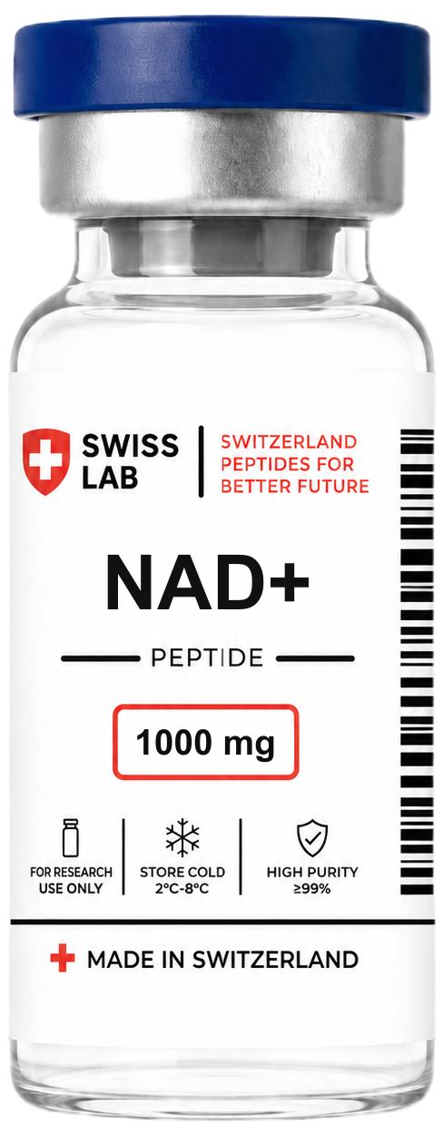
NAD+ — nicotinamide adenine dinucleotide — is one of the few molecules that appears in essentially every chapter of a biochemistry course. It is present in every living cell, it is the electron acceptor for most oxidative steps in catabolism, and it is simultaneously the raw material that a whole family of signalling enzymes chews through and throws away. Understanding it properly means holding both of those roles in mind at once.
This article is about the molecule and its chemistry: how NAD is built, what happens to it in a redox reaction, how it differs from NADP, which enzymes use it, and where a cell obtains it. A companion article on this site, the NAD+ research overview, covers NAD+ as a research compound and the sirtuin and aging-biology literature that surrounds it. Nothing here describes human use, supplementation, dosing, or therapeutic outcomes.
- NAD is a dinucleotide: a nicotinamide nucleotide and an adenine nucleotide joined through a pyrophosphate bridge.
- NAD+ is the oxidised form and acts as an electron acceptor; it takes a hydride ion at C4 of the nicotinamide ring and becomes NADH.
- It is a recycled cofactor in redox reactions but a genuinely consumed substrate for sirtuins, PARPs and CD38.
- NADP+ differs by a single phosphate on the 2′ position of the adenosine ribose, and is held in the opposite redox state.
- Most cells maintain their NAD+ through the salvage pathway from nicotinamide, not by building the ring from scratch.
NAD+ definition: what the molecule is in biology
NAD stands for nicotinamide adenine dinucleotide. It is a coenzyme — a small, non-protein organic molecule that an enzyme requires in order to carry out its reaction — with a molecular weight of about 663 g/mol. Its biological function is twofold. First, and best known, it is the universal electron carrier of catabolism: the molecule that collects the electrons stripped from sugars, fatty acids and amino acids and delivers them to the respiratory chain. Second, it is the ADP-ribose donor for a set of enzymes that use it up rather than recycle it, which is why cells must continually resynthesise it. The notation NAD+ refers specifically to the oxidised form and NADH to the reduced form; “NAD” without a suffix normally means the molecule irrespective of redox state.
The structure of NAD: two nucleotides and a pyrophosphate bridge
A nucleotide is a nitrogenous base plus a sugar plus a phosphate. NAD is two of them joined tail to tail. One half is nicotinamide mononucleotide (NMN): a nicotinamide group attached to D-ribose, carrying a 5′-phosphate. The other half is adenosine monophosphate (AMP): an adenine base attached to its own D-ribose, also carrying a 5′-phosphate. The two 5′-phosphates are linked to each other through a phosphoanhydride bond, giving the pyrophosphate (diphosphate) bridge that holds the molecule together. So the full inventory is one nicotinamide ring, one adenine ring, two ribose sugars in the β-furanose form, and two phosphates. Both bases are attached to their sugars by β-N-glycosidic bonds — adenine through its N9, nicotinamide through the nitrogen of its pyridine ring.
That last detail is the reason the nicotinamide ring is the business end of the molecule. Attaching the ring nitrogen to the sugar makes it a quaternary nitrogen with no lone pair left over, so it carries a formal positive charge. That is the “+” in NAD+, and it is what makes the pyridinium ring electron-poor and therefore a good electron acceptor. Everything on the other side of the pyrophosphate bridge — the adenine, the second ribose — takes no part in the chemistry at all. It functions as a molecular handle: a large, distinctive, negatively charged tail that enzymes grip, which is how a dehydrogenase tells NAD from any other small molecule floating past. The classic binding motif for that handle is the Rossmann fold, a βαβ unit with a glycine-rich loop found across hundreds of otherwise unrelated dehydrogenases.
The NAD+/NADH redox couple: NAD+ is the electron acceptor
Yes, NAD+ is an electron acceptor, and NAD+ is reduced to NADH. The transfer is a single chemical event: NAD+ accepts a hydride ion, written H−, which is one proton carrying two electrons with it, and that hydride adds to carbon 4 of the nicotinamide ring. When a dehydrogenase removes two hydrogen atoms from its substrate, only one of them travels as the hydride; the second is released into solution as a free proton. Hence the standard equation, substrate-H2 + NAD+ → substrate + NADH + H+, and hence the fact that the reduced form is written NADH rather than NADH2. Chemically the consequence is dramatic: the aromatic pyridinium ring becomes a non-aromatic 1,4-dihydropyridine, and the positive charge disappears. The standard reduction potential of the couple is about −0.32 V, which places NADH near the electron-rich end of the biological redox scale and is why it can reduce almost every downstream carrier it meets. Dehydrogenases are also stereospecific about the transfer, delivering the hydride to one particular face of C4 — the classic A-side and B-side distinction.
This is the point at which the common question “is NAD+ a substrate or a product?” usually arises, and the honest answer is that it is neither in the ordinary sense. It is a recycled cofactor, sometimes called a cosubstrate: it is written on the substrate side of one reaction and on the product side of the next, because it alternates between two forms rather than being made and destroyed. Glyceraldehyde-3-phosphate dehydrogenase turns NAD+ into NADH; lactate dehydrogenase and Complex I turn NADH back into NAD+. Across a pathway there is no net consumption, and a comparatively small pool — total cellular NAD is typically in the high-micromolar range — supports an enormous flux by turning over again and again. What complicates the picture is that a second class of enzymes really does consume it, cleaving the molecule apart rather than reducing it, so over a longer timescale NAD+ genuinely is a consumed substrate that has to be replaced.
One practical consequence of the ring chemistry deserves mention because it underpins a large fraction of all enzyme assays ever run. The 1,4-dihydropyridine ring of NADH absorbs ultraviolet light at around 340 nm with a molar extinction coefficient of roughly 6,220 M−1 cm−1, while the aromatic NAD+ ring does not absorb there at all. Both forms absorb at 260 nm because of the adenine, so 340 nm reports specifically on the reduced form. Any reaction that produces or consumes NADH can therefore be followed in real time as a rising or falling absorbance at 340 nm, and dehydrogenase reactions are routinely coupled to other enzymes purely to generate such a read-out. NADH is also fluorescent, exciting near 340 nm and emitting near 460 nm, whereas NAD+ is not, which extends the same trick to imaging.
NAD and NADP: one phosphate, two different jobs
NADP stands for nicotinamide adenine dinucleotide phosphate. Structurally it is NAD with one addition: a phosphate group esterified to the 2′-hydroxyl of the ribose on the adenosine half, installed by the enzyme NAD kinase. That phosphate sits far from carbon 4 and does not change the redox chemistry — NADP+ accepts a hydride in exactly the same way and becomes NADPH. What it changes is recognition. In many Rossmann-fold enzymes an aspartate residue hydrogen-bonds the free 2′- and 3′-hydroxyls of NAD; NADP-preferring enzymes replace that acidic residue with a basic one that accommodates the extra negative charge instead. One phosphate therefore sorts the two coenzymes into two almost non-overlapping enzyme populations, and the cell exploits that separation by holding them in opposite redox states. The NAD pool is kept strongly oxidised, so NAD+ vastly outnumbers NADH and there is always an acceptor available to keep catabolism running. The NADP pool is kept strongly reduced, so NADPH vastly outnumbers NADP+ and there is always a donor available for reductive chemistry.
| Property | NAD+ / NADH | NADP+ / NADPH |
|---|---|---|
| Structural difference | Free 2′-hydroxyl on the adenosine ribose | Phosphate esterified to that 2′-hydroxyl |
| Typical cellular state | Strongly oxidised: NAD+ greatly exceeds NADH | Strongly reduced: NADPH greatly exceeds NADP+ |
| Principal role | Catabolic oxidation — collects electrons and delivers them to the respiratory chain | Reductive biosynthesis and antioxidant chemistry — donates electrons |
| Where the reduced form is made | Glycolysis, the citric-acid cycle, beta-oxidation | Pentose phosphate pathway, malic enzyme, NADP-dependent isocitrate dehydrogenase |
| Representative enzymes | GAPDH, malate dehydrogenase, respiratory Complex I | Fatty acid synthase, glutathione reductase, thioredoxin reductase |
NAD-dependent enzymes: dehydrogenases and NAD+ consumers
The classic NAD-dependent enzymes are the dehydrogenases, and they are also the answer to where NADH is made. In glycolysis, glyceraldehyde-3-phosphate dehydrogenase reduces NAD+ to NADH in the cytosol. In the mitochondrial matrix the pyruvate dehydrogenase complex does it again, and the citric-acid cycle does it three more times per turn through isocitrate dehydrogenase, alpha-ketoglutarate dehydrogenase and malate dehydrogenase. Beta-oxidation of fatty acids contributes another NADH per cycle through 3-hydroxyacyl-CoA dehydrogenase, and amino-acid catabolism adds more through enzymes such as glutamate dehydrogenase. The electrons then leave: NADH is oxidised back to NAD+ by Complex I of the electron transport chain, NADH:ubiquinone oxidoreductase, which passes them to ubiquinone and uses the released energy to pump protons. Because the inner mitochondrial membrane will not let NADH across, cytosolic NADH reaches the chain indirectly through the malate–aspartate and glycerol-3-phosphate shuttles. When the chain cannot keep up, fermentative enzymes take over the regeneration instead: lactate dehydrogenase reoxidises cytosolic NADH so that glycolysis has NAD+ available and can continue.
The second group works on entirely different principles. Sirtuins, PARPs and the NAD+ glycohydrolases do not reduce NAD+; they break the N-glycosidic bond between the nicotinamide and its ribose, transfer the ADP-ribose portion somewhere useful, and release free nicotinamide. Sirtuins spend one NAD+ per protein deacylation, producing nicotinamide and 2′-O-acyl-ADP-ribose. PARPs transfer ADP-ribose units onto acceptor proteins, with PARP1 activation by DNA strand breaks being the most-studied case and one capable of drawing down the NAD+ pool substantially in cell models. CD38, CD157 and the TIR-domain enzyme SARM1 hydrolyse or cyclise NAD+ into ADP-ribose and cyclic ADP-ribose. Because all of these destroy the molecule, NAD+ turnover in a cell is fast — reported on a timescale of hours rather than days in several cell types — and the biosynthetic routes below are not a formality but a continuous requirement.
| Enzyme class | How NAD+ is used | Representative examples |
|---|---|---|
| NAD-dependent dehydrogenases | Recycled redox cofactor: NAD+ becomes NADH and back again, ring intact | GAPDH, lactate dehydrogenase, malate dehydrogenase, alcohol dehydrogenase |
| Respiratory Complex I | Reoxidises NADH to NAD+ and passes the electrons to ubiquinone | NADH:ubiquinone oxidoreductase |
| Sirtuins (SIRT1–7) | Consumed: cleaved during protein deacylation, releasing nicotinamide | SIRT1, SIRT3, SIRT6 |
| PARPs | Consumed: ADP-ribose transferred to acceptor proteins, nicotinamide released | PARP1, PARP2 |
| NAD+ glycohydrolases | Consumed: hydrolysed or cyclised to ADP-ribose and cyclic ADP-ribose | CD38, CD157, SARM1 |
Where does NAD+ come from? Three routes to the same molecule
NAD+ comes from three converging biosynthetic pathways, all of which end by building the dinucleotide from a mononucleotide intermediate. Only one of them constructs the pyridine ring from scratch; the other two start from a ring that has been eaten, absorbed or recovered. The convergence point is the enzyme family NMNAT (nicotinamide mononucleotide adenylyltransferase), which attaches the AMP half onto the nicotinamide half using ATP, and which exists as three isoforms with distinct addresses: NMNAT1 in the nucleus, NMNAT2 in the cytosol, NMNAT3 in mitochondria. That compartmentalisation matters, because NAD+ is a large charged molecule that does not cross membranes freely, so each compartment maintains its own pool.
- De novo synthesis from tryptophan, via the kynurenine pathway. Tryptophan is opened by IDO or TDO to N-formylkynurenine and processed through kynurenine and 3-hydroxyanthranilate to quinolinic acid, which quinolinate phosphoribosyltransferase converts to nicotinic acid mononucleotide. This is the only route that makes the ring itself, it runs mainly in liver and kidney, and it is inefficient — the classical textbook equivalence is roughly 60 mg of tryptophan for 1 mg of niacin.
- The Preiss–Handler pathway, from nicotinic acid. Nicotinic acid phosphoribosyltransferase condenses nicotinic acid with PRPP to give nicotinic acid mononucleotide, NMNAT adenylylates it to nicotinic acid adenine dinucleotide, and the glutamine-dependent NAD synthetase amidates that to NAD+.
- The salvage pathway, from nicotinamide. Nicotinamide phosphoribosyltransferase (NAMPT) condenses nicotinamide with PRPP to give nicotinamide mononucleotide, and NMNAT completes the molecule. NAMPT is the rate-limiting step, and its inhibition in cell culture collapses NAD+ levels within hours.
- Riboside and mononucleotide entry points. Nicotinamide riboside is phosphorylated by the nicotinamide riboside kinases NRK1 and NRK2 to nicotinamide mononucleotide, joining the salvage route one step downstream of NAMPT. Extracellular NMN is generally reported to be dephosphorylated by the ectoenzyme CD73 to the riboside before uptake; whether a direct NMN transporter also operates remains an open question in the literature.
In most mammalian cells the salvage pathway dominates, and the reason is structural rather than incidental. Every sirtuin, PARP and CD38 reaction releases one molecule of nicotinamide, so the cell is continuously generating exactly the substrate that NAMPT requires. Salvage closes that loop: nicotinamide out of the consuming enzymes, nicotinamide back into NMN and then NAD+. De novo synthesis from tryptophan supplies a comparatively small share outside the liver, and the Preiss–Handler route depends on nicotinic acid arriving from the diet or the gut microbiota. A cell deprived of salvage capacity cannot compensate through the other two routes quickly enough, which is why NAMPT inhibition is such a widely used experimental tool for depleting NAD+ in vitro.
What the evidence does not establish
Everything above is settled biochemistry, established across decades of enzymology and structural work, and it is worth being explicit that the marketing claims made about NAD+ products do not follow from it. That a molecule is essential to metabolism says nothing about what happens if you try to increase it, and the literature does not establish:
- Anti-aging, energy, or longevity outcomes in humans. Reports that tissue NAD+ declines with age come largely from animal models and are technically difficult to quantify, since NAD+ is labile and degrades during extraction; they are not evidence of a human benefit from raising it.
- That raising a measured NAD+ metabolite level produces any functional outcome. Trials of precursor molecules have generally used biochemical markers as endpoints, not clinical ones.
- That NAD+ supplied from outside a cell reaches the intracellular pool intact. NAD+ is large and charged and does not cross the plasma membrane freely; extracellular NAD+ is largely degraded by ectoenzymes before anything is taken up.
- Human dosing, routes of administration, or safety — none of these are addressed by the biochemistry described here.
- That the sirtuin and aging-biology findings in yeast, worms, flies and mice scale to people. This remains unknown.
The references below point to the primary literature on NAD biosynthesis, redox enzymology and the NAD+-consuming enzyme families, and the product page lists the analytical documentation available for the research material.
Frequently asked questions
- Is NAD+ a substrate or a product?
- Both, depending on the reaction, and strictly speaking neither. NAD+ is a recycled cofactor: a dehydrogenase writes it on the substrate side and produces NADH, while Complex I or lactate dehydrogenase does the reverse. It is not consumed by redox chemistry. It is, however, a genuinely consumed substrate for sirtuins, PARPs and CD38, which cleave it and release nicotinamide.
- Is NAD an electron acceptor, and what is NAD reduced to?
- NAD+ is the oxidised form and acts as an electron acceptor. It takes on a hydride ion — one proton plus two electrons — at carbon 4 of the nicotinamide ring and is reduced to NADH, with the second hydrogen from the substrate released into solution as a free proton. NADH is the electron donor of the pair and hands its electrons to Complex I of the electron transport chain.
- Where does NAD+ come from and where is NADH made?
- NAD+ is synthesised by three routes: de novo from tryptophan via the kynurenine pathway, the Preiss–Handler pathway from nicotinic acid, and the salvage pathway from nicotinamide via NAMPT, which dominates in most cells. NADH is made wherever a dehydrogenase reduces NAD+ — principally in glycolysis in the cytosol, and in the pyruvate dehydrogenase complex, the citric-acid cycle and beta-oxidation in mitochondria.
- What is the difference between NAD and NADP?
- A single phosphate group esterified to the 2′-hydroxyl of the adenosine ribose. It does not alter the redox chemistry but it changes which enzymes bind the coenzyme, and the cell holds the two pools in opposite states: NAD mostly oxidised for catabolism, NADP mostly reduced for biosynthesis and antioxidant defence.
- Why does NADH absorb at 340 nm?
- Because reduction converts the aromatic pyridinium ring of NAD+ into a non-aromatic 1,4-dihydropyridine, which has an absorbance maximum near 340 nm with a molar extinction coefficient of about 6,220 M−1 cm−1. NAD+ does not absorb there, so the change at 340 nm reports specifically on the reduced form and is the basis of most dehydrogenase assays.